The structure of the human eye is similar to an optical camera. The function of the retina, optic nerve, and visual center of the brain is the film of the camera; iris is the aperture, and cornea is the lens of the camera. The cornea is to be turbid what the camera lens is to be worn, which will affect the image quality. Keratopathy is the most common eye disease, which can make the transparent cornea appear gray and turbid, block the light into the eye, and finally cause blurred vision, decline, and even blindness.

If you replace the worn lens with a good one, you still can take a clear picture, which is the same as what we usually call corneal transplantation. In the past, corneas used in keratoplasty were donated by others. However, because of the extremely low corneal donation rate, only one thousandth of the blind patients with corneal diseases undergo corneal transplantation in China. In this case, artificial cornea transplantation is taken into the public’s eyes.
Not long ago, Israeli scientists successfully implanted artificial cornea into an elderly patient who had been blind for ten years, enabling him to recover his vision almost immediately after the operation. The success of artificial cornea transplantation by Israeli scientists is a major medical breakthrough, which will make thousands of blind patients see the light again.

As a matter of fact, keratoprosthesis is not regarded as a complicated technical work but with few applications in the world because of various uncontrollable factors, such as poor biocompatibility and side effects. It is undeniable that this success will make future practice more meaningful and confident though artificial cornea transplantation is still in the experimental stage.
The careful research and clinical trials of ophthalmologists at Rabin Medical Center contribute to the success of this operation. Moreover, various ophthalmic equipment also made their efforts in the process. For example, a slit lamp microscope provides doctors with high-definition images and cornea analysis before and after surgery.
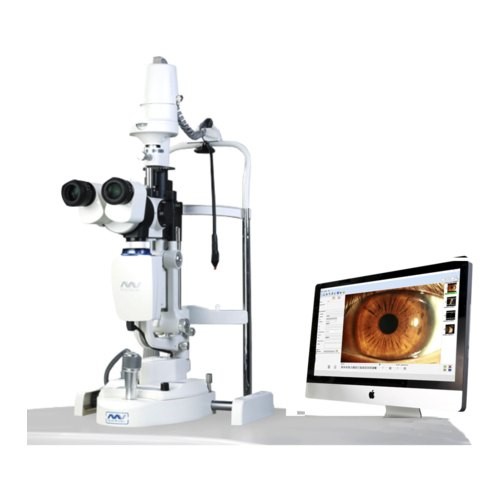
A digital slit lamp is a microscope with bright light used in the ophthalmic examination. It enables ophthalmologists to observe the different structures of the eyes more carefully so as to determine the critical schemas for eye health and detection of eye diseases.
Before corneal transplantation, a slit lamp microscope, intraocular pressure measurement, and ophthalmic imaging system will be used by ophthalmologists for clinical evaluation of implanted eyes. If abnormal substances accumulate in the cornea during slit lamp examination, it may be a symptom of corneal dystrophy, which will lead to blurred vision and loss of vision without timely treatment. With a slit lamp microscope, ophthalmologists are good at determining the operation plan according to the special situation of the patient.
With the extensive use of keratoplasty, to visualize corneal thickness and perceive various corneal depths during surgery become inevitable steps for corneal surgeons. The slit view provided by the digital slit lamp allows ophthalmologists to directly view the corneal thickness in real-time and promote the smooth operation of corneal transplantation.
Given that artificial cornea is made of a synthetic and non-degradable porous material, the middle of which is a transparent lens, sutures is what it needs to fix artificial cornea on the surface of the eye. Therefore, the digital slit lamp still plays a role in determining the retention rate of the artificial cornea after surgery; furthermore, whether it has poor biocompatibility.
It is necessary to follow up with the patients after keratoplasty. Slit lamp microscope with a built-in yellow filter and cobalt blue light is the right hand for doctors to observe the healing of the incision and provide timely solutions.
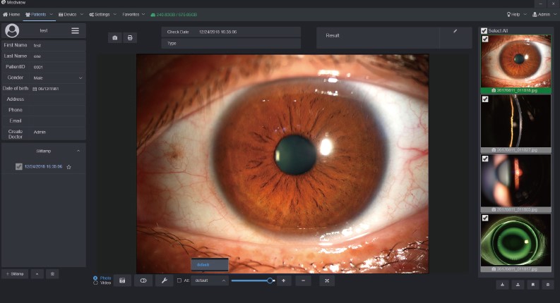
A slit lamp microscope is so crucial to corneal transplantation that even Professor Irit Bahar, director of Ophthalmology at Rabin Medical Center in Israel, used MediWorks slit lamp to perform an operation. Our slit lamp microscope occupies extraordinary resolution and images analysis function to help ophthalmologists with higher accuracy and safe diagnosis results.
In addition, personalized functions such as patient management systems and custom auto-exposure value settings can make doctors more flexible in diagnosing ophthalmic diseases. If you are interested in MediWorks slit lamp, don't hesitate to contact us. We always give priority to the customer's needs.