The team of Dr. Steven L Maskin, from Dry Eye and Cornea Treatment Center, Tampa, FL. in the United States, published their latest research titled Meibography Guided Intraductal Meibomian Gland Probing Using Real-Time Infrared Video Feed on CLINICAL SCIENCE.
The main purpose of this study was to describe and quantify the findings of intraductal obstruction during the detection of expressible and non-expressible meibomian glands (MGS) in patients with obstructive meibomian gland dysfunction by using a 1-mm intraductal MG probe.
Obstructive meibomian gland dysfunction (o-MGD) is considered the most common cause of dry eye in the world. If untreated, o-MGD can cause inflammation of the eyelid and ocular surface, followed by infrared meibomian imaging, suggesting meibomian gland (MG) atrophy. The traditional explanation and treatment of glandular obstruction mainly focus on thick tarsal thickening or keratotic lumen fragments. However, the results are inconsistent and often not enough to cause dissatisfaction and depression of patients and physicians.
In order to better understand the disease, Dr. Steven L Maskin developed the meibomian gland probing (MGP) technique to detect obstructive MG that is intolerable to traditional treatment. They performed probe detection of 996 glandular foramens in 38 patients and recorded real-time images using a high-definition slit lamp with a unique infrared light source module for the retrospective study.
The slit lamp is a microscope with bright light used in the ophthalmic examination. As a key tool to determine eye health and detect eye diseases, it enables ophthalmologists to observe the different structures in the eyes more carefully. A slit lamp with a unique infrared light source module can realize the visualization of intraductal probing for the meibomian gland, microtubule therapy injection, and palpebral fat sample retrieval in the experiment, which provides safety support for clinical technology.
In this experiment, MediWorks S390L Firefly WDR gave Dr. Steven L Maskin a hand to realize real-time imaging of infrared video. MediWorks S390L slit lamp also exerts great influences on the conclusion from Dr. Steven L Maskin, which is in addition to topical anesthetic ointment, the use of Maskin probe and catheter especially designed and sized for meibomian gland will greatly improve the comfort of patients. Moreover, it is necessary for symptomatic patients with MGD and asymptomatic patients with subclinical diseases to confirm the diagnosis of meibomian gland deficiency by slit lamp with an infrared light source module.
The visualization of MediWorks S390L Firefly WDR makes it possible to search the whole or local gland treatment and tibia specimens, which greatly promotes future research, treatment, and regeneration opportunities. In addition to this, the built-in WDR and firefly optical system can make the ophthalmologist's diagnosis more accurate.
Its intelligent case management system supports multiple case comparison function so that the effect of follow-up is clear at a glance. The system also supports a DICOM port, which can easily connect with the medical system of the hospital. Finally, it is worth mentioning that the built-in yellow filter of MediWorks S390L Firefly WDR can increase the contrast of corneal fluorescein sodium staining image so as to improve the positive detection rate of early corneal epithelial staining.
IR video feed with colour slit lamp imaging was obtained using the Mediworks S390L WDR FireFly Digital Slit Lamp from Eyefficient. This technology enabled visualisation of intraductal probing, microtube therapeutic injection and meibum specimen retrieval. Parameters evaluated were percentage of total probed glands with a visualised intraductal probe beyond the orifice, detection of any false passage and correlation of probe findings revealing specific gland intraductal space characteristics with corresponding meibography images. Probes and microtubes were obtained from Katena Products. All procedures adhered to the principles of the Declaration of Helsinki, and all patients provided informed consent. This study received an independent review board exempt review determination by an independent review board.
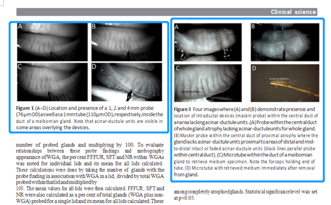
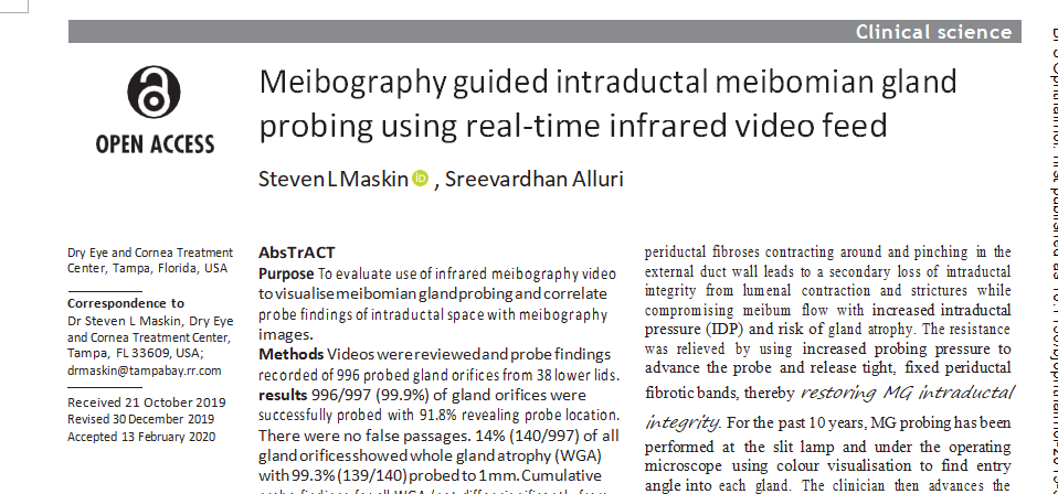
Figure 1(A–D) Location and presence of a 1, 2 and 4 mm probe
(76 μm OD) as well as a 1 mm tube (110 μm OD), respectively, inside the duct of a meibomian gland. Note that acinar-ductule units are visible in some areas overlying the devices.
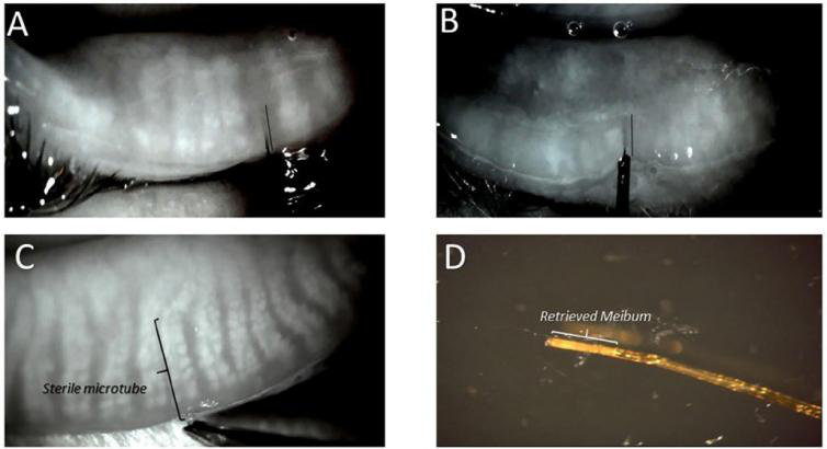
Figure 2 Four images where (A) and (B) demonstrate presence and location of intraductal devices (maskin probe) within the central duct of an area lacking acinar-ductule units. (A) Probe within the central duct of whole gland atrophy lacking acinar-ductule units for whole gland.(B) Maskin probe within the central duct of proximal atrophy where the gland lacks acinar-ductule units proximal to areas of distal and mid-to-distal intact or faded acinar-ductule units (black lines parallel probe within central duct). (C) Microtube within the duct of a meibomian gland to retrieve meibum specimen. Note the forceps holding end of tube. (D) Microtube with retrieved meibum immediately after removal from gland.
Attention should be paid to the annual examination of meibomian gland morphology to maintain its integrity. And it is wise for medical institutions to use advanced slit lamp because it can not only improve the accuracy of diagnosis but also directly improve work efficiency. As one of the major ophthalmic instrument providers with advanced R & D apartments, MediWorks is committed to providing high-quality ophthalmic instruments for thousands of ophthalmic clinics at home and abroad by combining optical, mechanical, electronic, and other cutting-edge technologies.
Original download resource for reference, please refer to :https://bjo.bmj.com/content/104/12/1676